Research Areas and Projects


Identification of candidate anti-cancer molecules

Recent WHO-IARC data reports that cancer is the major cause of morbidity and mortality, with approximately 14 million new cases and 8 million cancer-related deaths in 2012 (GLOBOCAN-2014). Traditional chemotherapeutic agents remain inefficient for the treatment of cancer with poor prognosis. Therefore there is a need for targeted therapies. Our group has been working on the molecular biology of liver cancer with a specific aim of discovery of novel candidate molecules for the treatment of this deadly Disease. Our focus is liver cancer, which is the 5th most common and 3rd lethal cancer and associated with poor overall survival. Major molecular and cellular biology approaches

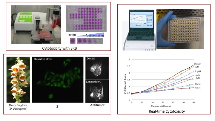
  1. NCI-60 Cytotoxicity screening of small molecules on liver, colon and breast cancer cells
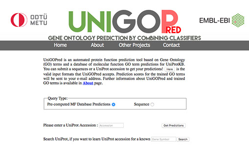
  2. Evaluation of cytotoxicities in real time with RT-CES systems
  3. Investigation of the induced morphological changes
  4. Cell cycle experiments
  5. Wound healing and migration assay with time lapse camera
  6. Characterization of cell death mechanism
  7. Mice xenograft experiments
We collaborate with various national and international institutions on molecular cellular biology and bioinformatics of cancer cells. Our group is an active member of COST action CM1106 (StemChem). We have also collaborations on protein function annotation with UniProt- EBI and with Fraunhofer Institute for cancer stem cell image analysis. We tested more than 2500 novel compounds for their cytotoxicity. Molecules with IC50 values less than 10µM have been further analyzed and were reported.

Responsive image

Protein Function Prediction

UniGOPred is an automated protein function prediction tool and GO prediction database of UniProtKB for all three categories of GO. It is composed of two components:

  1. A novel prediction tool for sequence based GO annotations.
  2. A database of pre-computed MF GO term predictions for UniProtKB entries and a database of pre-computed BP and CC GO term predictions for Model Organisms.
List of model organims are obtained from NIH website and the sequences for the corresponding model organisms are obtained from UniProtKB. The main characteristics that differentiate our approach from existing studies are the use of a combination of independent classifiers, and novel data preparation and evaluation methods. UniGOPred is trained for 514 Molecular Function GO terms, 438 Cellular Component GO terms and 2909 Biological Process GO terms. When a protein sequence is given as input, the predictions are sent to the e-mail address of the user. Alternatively, a UniProt accession can be submitted to get pre-computed MF predictions as an e-mail.

Responsive image

MAPK/PI3K/AKT pathway in Liver cancer

We have constructed a detailed map of AKT-signaling pathway by compiling the data in around 498 publications. Currently, the pathway has 254 proteins and 478 connections with inhibition, activation or just binding edge properties in Cytoscape. Sequences and available PDB structures have been mapped to the proteins in AKT signaling pathway and ready to be used in the next step. PI3K/AKT/MTOR cell survival signaling is frequently altered in cancer cells through genetic or epigenetic modifications. Therefore this pathway is considered as a good candidate for targeted drug discovery. Our wet lab studies mainly focuses on the screening of the small molecules on AKT-signaling pathway and in vitro validation of resulting target set of proteins and in vivo xenograft validation of anti cancer activities Laboratory work

  1. in vitro bioactivity screening of the predicted small molecules.
  2. AKT signaling analysis and interactome mapping.
  3. RNAseq and microarray experiments done with the inhibitors of AKT pathway.

Responsive image

Computer assisted microscopy

Computerized microscopy-based image processing is a key technology for determining the information relevant for diagnosis and prognosis of various diseases including cancer. Tool based on the determination of tumor heterogeneity ratio in histopathological images may enhance cancer diagnosis prognosis and treatment approaches. By using an automated microscopy and image processing system, high differential diagnostic accuracy can be achieved, while human factors such as subjectiveness and workload can be reduced. The required high–throughput acquisition of microscopic images from pathological tissue samples can be done e.g. by automating conventional microscopes (which is usually error-prone and costly) or by the use of commercially available slide scanners (which are usually closed systems and rather expensive).

In collaboration with Dr. Enis Cetin, Dr. Aysegül Uner, Cigdem Gunduz Demir and Dr. Christian Münzenmayer goal is to develop new methods and systems to improve diagnostic and prognostic techniques in cancer treatment by computer-assisted microscopy.

Responsive image
Informatics Institute